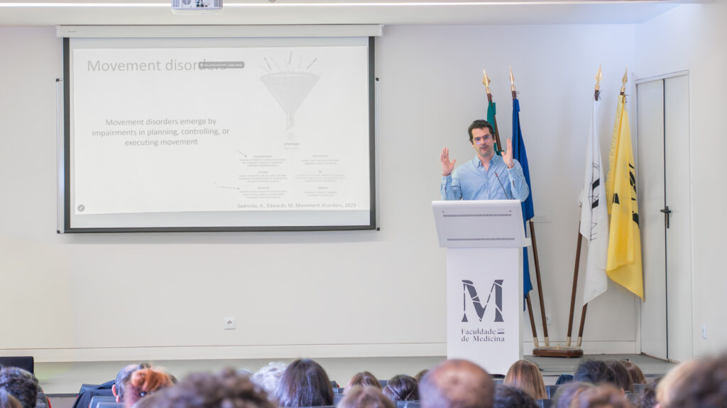
Movement disorders emerge when brain circuits that normally coordinate voluntary action become imbalanced. In conditions such as Parkinson’s disease, the degeneration of dopamine-sensitive pathways alters how movements are initiated and controlled, revealing how deeply motor behaviour depends on finely tuned neural communication. Research in this field increasingly focuses on identifying which circuits fail first and how their dysfunction produces specific symptoms. Why does a movement begin smoothly in a healthy brain, but becomes delayed, fragmented, or involuntary when neural circuits fail? That is the question scientists, like the psychiatrist and researcher at Champalimaud Foundation Joaquim Alves da Silva, have been trying to answer to.
During his visit to GIMM, Joaquim gave a lesson to the PhD students and presented a seminar on how neural circuits control movement and how their dysfunction gives rise to movement disorders, focusing on Parkinson’s disease and dystonia.
The talk began by highlighting the remarkable complexity of everyday motor actions, like walking, and how these rely on a finely coordinated interplay between cortical and subcortical brain regions, particularly the basal ganglia, cerebellum and motor thalamus, with Joaquim noting that movement disorders offer a unique window into understanding these circuits, since different diseases often produce distinct and reproducible motor symptoms despite diverse underlying causes.
A significant part of the seminar focused on Parkinson’s disease and on the role of dopamine neurons in movement initiation. Drawing on previous and ongoing work in mouse models, he showed how dopamine activity is closely linked to the probability of initiating movement and to movement vigor, especially before movement begins. His lab has demonstrated that transient inhibition of dopamine neurons can acutely reduce spontaneous movement initiation, reproducing features of bradykinesia, a hallmark symptom of the disease.
One of the central questions addressed by his current research is why externally cued movements, such as walking over visual markers or responding to an auditory signal, can temporarily improve motor performance in Parkinsonian patients, even when self-initiated movement remains impaired. To investigate this, the lab has developed behavioral paradigms in mice that directly compare self-paced and cued locomotion. Preliminary results suggest that external cues may recruit alternative neural pathways that bypass dysfunctional basal ganglia output.
This work is now being extended through a project funded by the European Research Council, combining progressive mouse models of Parkinson’s disease, optogenetics, electrophysiology and viral tracing to identify how different motor circuits converge in the motor thalamus during movement initiation.
In the second part of the seminar, the topic was dystonia, a hyperkinetic movement disorder characterized by involuntary muscle contractions and abnormal postures. Joaquim discussed the challenge of understanding dystonia at circuit level, particularly because many genetic mouse models do not show obvious spontaneous symptoms.
To address this, his group is developing new experimental platforms that overtrain mice in highly repetitive, precision-based reaching tasks, inspired by task-specific dystonias such as writer’s cramp and musician’s dystonia. These systems allow continuous behavioral monitoring and muscle recordings, with the goal of detecting subtle abnormalities in motor control and muscle coordination that may reveal early circuit dysfunction.
By studying these disease models, scientists expect to provide both clinically relevant insights and a deeper understanding of the fundamental neural mechanisms underlying motor control.