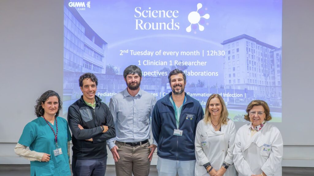
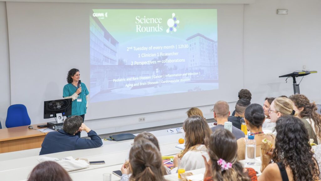
On May 12 we have had a session on in interstitial lung diseases, by João Eusébio, MD, Pneumology Department, ULSSM, and GIMM’s Group Leader Pavel Hanč, on “Nociceptors and tumor immunity
Bronchoalveolar biomarkers and the future of interstitial lung disease diagnosis
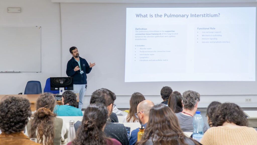
Few things are as terrifying as being unable to breathe. Interstitial lung diseases (ILD) affect the microscopic space between the alveolar epithelium and the blood vessels (the so-called interstitium) impairing the lungs’ ability to exchange oxygen. More than 200 diseases fall under the umbrella of ILDs, a complex and often difficult-to-diagnose group of disorders that affect the lung’s internal architecture. In the talk “Evaluation of endoscopic and bronchoalveolar biomarkers in interstitial lung diseases”, João Eusébio,MD (Pneumology Department, ULSSM),explored how bronchoscopy and bronchoalveolar lavage (BAL) are helping doctors better understand these conditions and potentially come up with more precise, personalised treatments.
At the centre of the talk was this question: how can doctors obtain clearer biological signals from the lungs themselves?
João Eusébio started by explaining that the term “interstitial” can be misleading. “These diseases not only affect the interstitium itself, but also the epithelial and endothelial compartments of the lung,” he noted, highlighting the complexity of conditions that range from inflammatory disorders to progressive fibrosis.
Despite major advances in imaging and pathology, diagnosing ILDs remains challenging. Different diseases frequently share symptoms, CT scan patterns and even histopathological characteristics resulting in around a quarter of patients still not receiving a definitive diagnosis. “Different diseases share clinical features, so we need biomarkers to tell them apart,” he explained.
That is where bronchoscopy, and specifically bronchoalveolar lavage (BAL), becomes particularly valuable. BAL is a minimally invasive procedure in which saline is introduced into a targeted region of the lung and then retrieved for analysis, allowing clinicians to study the local immune and fibrotic microenvironment directly.
“It’s really safe to do, repeatable, and it reflects the alveolar immune microenvironment,” he said.
While current clinical practice mainly focuses on analysing the cellular composition of BAL samples, João Eusébio highlighted how much more information may still be hidden within them. Researchers can already identify distinct immune patterns associated with different diseases. Lymphocyte-rich BAL samples, for example, are commonly associated with hypersensitivity pneumonitis and sarcoidosis, while neutrophil predominance is linked to progressive fibrosis and poorer outcomes in idiopathic pulmonary fibrosis.
“The more inflamed you are, the more lymphocytes you have,” he explained, noting that inflammatory profiles may also predict better responses to immunosuppressive therapies.
Macrophages, meanwhile, emerged as key players in fibrosis progression. Activated macrophages promote extracellular matrix deposition and fibroblast activation, contributing to the scarring process that progressively damages lung function.
Beyond cellular analysis, the field is rapidly expanding into molecular biomarkers and “omics” technologies. Researchers are investigating cytokines, chemokines, extracellular vesicles, transcriptomics and proteomics extracted from BAL samples, searching for signatures that could improve diagnosis, predict disease progression, or guide treatment selection.
“There’s a lot of new biomarkers that we can look at,” he said. “They all show something, but we still haven’t been able to use them clinically.”
One of the major challenges remains standardisation. Variability in sampling techniques, assay methods and validation thresholds continues to limit the translation of many promising biomarkers into routine care. The integration of imaging, BAL cellularity, molecular biomarkers and genomic data into multimodal AI-driven models could eventually transform how ILDs are classified and treated.
“We’re going from clinical entities toward biological and physiopathological classifications,” Eusébio said, highlighting the research opportunities emerging from this field. With access to high clinical volumes, bronchoscopy expertise and the possibility of establishing BAL biobanks, the department hopes to stimulate further translational research capable of bridging laboratory discoveries with clinical practice. So, although interstitial lung diseases are highly heterogeneous, many eventually converge toward fibrosis. In general, these diseases tend to evolve from an initial inflammatory state into a fibrotic stage marked by progressive and often irreversible scarring of the lung tissue. Identifying the disease while inflammation still predominates is therefore critical, since at this stage patients may still benefit from treatments capable of controlling – or even correcting – the pathological process. Once fibrosis becomes established, however, therapeutic options become far more limited. Understanding and predicting that transition may depend on looking more closely into the lung itself. Finding biomarkers capable of identifying which patients and when move from an inflammatory to a fibrotic state could significantly change clinical outcomes.
Exploring the hidden dialogue between nociceptors and cancer
Pain has long been understood as one of the defining signs of inflammation. But what if pain-sensing neurons are not merely passive messengers warning the body of danger? What if they actively shape immune responses and even help tumours evade destruction?
That was the central question explored in Pavel Hanč talk, “Nociceptors drive immunosuppressive monocyte differentiation to prevent tumor immunity”,on neuro-immune interactions in cancer biology, where researchers presented emerging evidence that nociceptors – the specialised neurons responsible for detecting harmful stimuli – may play an active role in tumour progression.
“I like to start my talks with a reminder that just because we’ve known something forever doesn’t mean that’s where the story ends,” Pavel noted.
Nociceptors, derived from the Latin nocere (“to harm”), are best known for transmitting pain signals from tissues to the spinal cord and brain. But beyond their sensory function, these neurons also release neuropeptides and signalling molecules directly into tissues. Many of these molecules possess strong immunomodulatory properties.
This raises a fundamental biological question: why would neurons regulate immunity?
The answer, Pavel argued, lies in evolution. Both nociceptors and the immune system exist to protect the organism from harm, but they do so in complementary ways. Nociceptors react rapidly to tissue stress or injury, while immune cells provide specialised and sustained defensive responses.
“Nociceptors should really serve as sensors or controllers here, and the immune system as the effector,” he explained.
In ideal circumstances, this communication helps coordinate tissue protection and repair. But in diseases such as asthma, chronic inflammation, infection and cancer these interactions can become maladaptive. And Pavel presented the example of bladder cancer, where researchers uncovered evidence that nociceptors may actively help tumours escape immune attack.
Using mouse models of bladder cancer, the team recreated a phenomenon known as intraluminal seeding, where tumour cells released into the bladder can re-establish themselves in previously healthy tissue. Once implanted, these tumours became densely innervated by nociceptor fibres and heavily infiltrated by immune cells.

But one immune population stood out in particular: myeloid-derived suppressor cells (MDSCs), highly immunosuppressive cells capable of blocking anti-tumour T-cell responses.
“What we found is that our T-cells are excluded from the tumour,” he explained. “They’re sitting behind what almost looks like a wall of these suppressive myeloid cells.”
To test whether nociceptors were involved in building this immunosuppressive environment, the researchers chemically eliminated pain-sensing neurons in mice before tumour implantation. The results were clear. In most animals lacking nociceptors, tumours failed to establish themselves. Instead, researchers observed only scar-like fibrotic tissue and evidence of tumour cell destruction. This protective effect disappeared in mice lacking adaptive immune cells, confirming that tumour rejection depended on functional T-cell responses.
The findings suggested that nociceptors were not simply present near tumours, but were actively shaping the tumour microenvironment in ways that suppressed immunity.
Further experiments revealed that nociceptors directly interacted with incoming monocytes, pushing them toward an immunosuppressive MDSC-like state. These altered immune cells not only inhibited T-cell activity, but also began proliferating themselves, amplifying immune suppression inside the tumour.
Transcriptomic analyses showed that monocytes exposed to nociceptors in vitro became molecularly similar to suppressive cells isolated directly from tumours.
“It’s not just that you end up with a phenotypic shift,” the speaker noted. “These immunosuppressive cells also begin to proliferate.”
The communication mechanism itself remains under investigation, although early evidence suggests it requires direct cell-to-cell contact and involves specific transcription factors such as SP1.
Taken together, the work proposes a new model of tumour immune evasion: as bladder tumour cells begin to establish themselves, they activate nearby nociceptors, which in turn reprogram monocytes into suppressive cells that shield the tumour from T-cell attack.
Without nociceptors, however, this protective barrier never forms, allowing the immune system to eliminate the tumour before it fully develops.
Beyond bladder cancer, the research contributes to a rapidly expanding field investigating how the nervous system shapes immunity across cancer, inflammation and tissue repair. What was once considered a simple pain pathway is increasingly emerging as a central regulator of immune biology. The findings also resonate with broader questions raised in other areas of inflammatory disease research, including interstitial lung diseases discussed. If nociceptors actively shape immune responses and tissue microenvironments, they may also influence how inflammation progresses, or fails to resolve, in fibrotic diseases. These emerging connections between the nervous system, immunity and tissue remodelling point toward a more integrated view of disease, where neurons, immune cells and damaged tissues continuously communicate to determine clinical outcomes.